

High-Yield Pathology for NBCE Part I: The General Pathology You Must Know
Pathology can feel like the biggest subject on Part I. Every organ system, every disease, every buzzword. It sits in Session 2 between Chemistry and Microbiology, and plenty of students treat it as a giant list to memorize.
There's a better way in. Most pathology questions, even organ-specific ones, rest on a small set of general principles: how cells respond to stress, how they die, how the body responds with inflammation, how tumors behave, and how clots form. Learn those well, and organ pathology becomes those same ideas applied to different tissues.
This guide covers that foundation, with tables you can review in minutes and practice questions at the end.
New to Part I prep? Start with our 10-week NBCE Part I study plan.
Cell Adaptation and Injury: The Continuum
Cells respond to stress in a predictable order. First they adapt. If the stress is too much, they're injured. If it continues past a certain point, they die.
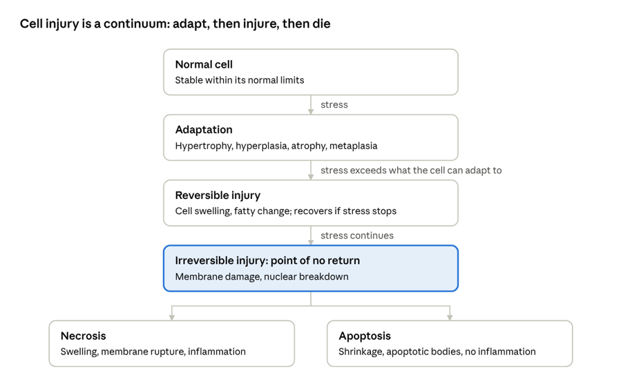
cell response to stress · adaptation to cell death
The four adaptations are a favorite source of definition questions, so know them precisely:
|
Adaptation |
What changes |
Classic example |
|
Hypertrophy |
Cells get bigger |
Heart muscle in long-standing hypertension |
|
Hyperplasia |
Cells increase in number |
Endometrium during the menstrual cycle; benign prostatic hyperplasia |
|
Atrophy |
Cells shrink |
Muscle wasting after immobilization |
|
Metaplasia |
One mature cell type replaced by another |
Barrett esophagus (squamous to columnar); smoker's airway (columnar to squamous) |
Watch the trap: dysplasia isn't an adaptation. It's disordered growth with abnormal cells, and it's considered precancerous.
The 6 Types of Necrosis
Necrosis questions almost always give you a tissue or a cause and ask for the type. Learn each type by its setting.
|
Type |
What it looks like |
Classic setting |
|
Coagulative |
Tissue outline preserved, but cells are dead |
Ischemia in most solid organs, like a heart attack (myocardial infarction) |
|
Liquefactive |
Tissue digested into liquid |
Brain infarcts; abscesses |
|
Caseous |
Soft, "cheese-like" material |
Tuberculosis |
|
Fat |
Chalky white deposits (fat combines with calcium) |
Acute pancreatitis |
|
Fibrinoid |
Pink, fibrin-like material in vessel walls |
Vasculitis; malignant hypertension |
|
Gangrenous |
Coagulative necrosis of a limb, sometimes with infection |
Lower limb ischemia, often in diabetes |
Memory hook: ischemia causes coagulative necrosis everywhere except the brain. The brain liquefies.
Necrosis vs Apoptosis
|
|
Necrosis |
Apoptosis |
|
Trigger |
Always pathologic |
Can be normal (development, cell turnover) or pathologic |
|
Cell size |
Swells |
Shrinks |
|
Membrane |
Ruptures |
Stays intact |
|
Inflammation |
Yes |
No |
|
Key players |
Loss of ATP, membrane damage |
Caspases; cell breaks into apoptotic bodies |
Acute vs Chronic Inflammation
The question usually hinges on the cell type. If you remember which cells show up when, you can answer most inflammation questions.
|
|
Acute inflammation |
Chronic inflammation |
|
Timing |
Minutes to days |
Weeks to years |
|
Main cells |
Neutrophils |
Macrophages, lymphocytes, plasma cells |
|
Classic signs |
Redness, heat, swelling, pain, loss of function |
Tissue destruction alongside repair and scarring (fibrosis) |
|
Typical causes |
Infection, trauma, tissue death |
Persistent infection, autoimmune disease, long-term irritants |
|
Possible outcomes |
Full healing, abscess, or progression to chronic |
Fibrosis; sometimes granulomas |
Granulomas are a special pattern of chronic inflammation: clusters of activated macrophages (epithelioid cells), often with giant cells. The classic contrast:
- Caseating granulomas: tuberculosis
- Non-caseating granulomas: sarcoidosis
The classic Latin names for the cardinal signs are rubor (redness), calor (heat), tumor (swelling), dolor (pain) and functio laesa (loss of function). Expect to see them in questions.
Neoplasia: How Tumors Are Named and Judged
Naming rules come up constantly:
- "-oma" usually means benign (lipoma, adenoma, leiomyoma).
- Carcinoma is a malignant tumor of epithelial cells. Sarcoma is a malignant tumor of connective tissue (mesenchymal) cells.
- The exceptions are malignant despite the "-oma": lymphoma, melanoma and mesothelioma.
|
|
Benign |
Malignant |
|
Growth |
Slow, well-defined borders |
Often fast, invades nearby tissue |
|
Differentiation |
Looks like the tissue of origin |
Ranges from well to poorly differentiated |
|
Metastasis |
Never |
Can spread |
Grade vs stage: grade describes how abnormal the cells look under the microscope. Stage describes how far the tumor has spread. Stage usually matters more for prognosis.
How malignant tumors spread: carcinomas classically spread through lymphatics, and sarcomas through the blood. There are exceptions, but this is the default answer.
Hemodynamics: Clots and Swelling
Virchow's triad lists the three factors that promote a blood clot (thrombosis):
- Stasis of blood flow, like immobility or long flights
- Endothelial injury, like trauma or atherosclerosis
- Hypercoagulability, like pregnancy or certain genetic conditions
The most common source of a pulmonary embolism is a deep vein thrombosis in the legs.
Edema has four basic causes: increased hydrostatic pressure (heart failure), decreased oncotic pressure (low albumin from liver or kidney disease), increased vessel permeability (inflammation) and lymphatic obstruction.
Test Yourself: 5 Practice Questions
Cover the answers, commit to a choice, then check.
- A patient dies several days after a stroke. What type of necrosis would you expect in the affected brain tissue? A. Coagulative B. Liquefactive C. Caseous D. Fat
- In a long-term smoker, the columnar cells lining the bronchi are replaced by squamous cells. What is this called? A. Hyperplasia B. Dysplasia C. Metaplasia D. Hypertrophy
- Which cell type dominates in acute inflammation? A. Neutrophils B. Lymphocytes C. Plasma cells D. Macrophages
- Which of these tumors is malignant? A. Lipoma B. Adenoma C. Melanoma D. Leiomyoma
- A patient develops a deep vein thrombosis after a 14-hour flight. Which part of Virchow's triad best explains it? A. Endothelial injury B. Stasis C. Hypercoagulability D. Increased oncotic pressure
Answers
- Ischemia causes coagulative necrosis everywhere except the brain, which undergoes liquefactive necrosis.
- One mature cell type replaced by another is metaplasia.
- Neutrophils lead acute inflammation; macrophages and lymphocytes take over in chronic inflammation.
- Melanoma is one of the malignant "-omas," along with lymphoma and mesothelioma.
- Sitting still for hours slows blood flow in the legs.
Missed one? Add it to your miss log, as we describe in our Part I study plan.
Pathology FAQs
How should I study pathology for NBCE Part I? Start with general pathology (cell injury, necrosis, inflammation, neoplasia and hemodynamics), then move into organ systems. The general principles make organ pathology much easier to learn.
What's the difference between necrosis and apoptosis? Necrosis is always pathologic: cells swell, burst and trigger inflammation. Apoptosis is programmed cell death: cells shrink, break into apoptotic bodies and cause no inflammation.
What's the difference between metaplasia and dysplasia? Metaplasia is one normal, mature cell type replacing another and can be reversible. Dysplasia is disordered growth of abnormal cells and is considered precancerous.
When is Pathology tested on NBCE Part I? In Session 2, between Chemistry and Microbiology. See the NBCE Part I exam pagefor current details.
What else should I review for Session 2? Our biochem pathways guide and microbiology cheat sheet cover the other two Session 2 domains.
Make Pathology Manageable
General pathology is the foundation. Organ-system pathology is where the volume is, and that's where most students lose time trying to figure out what's high-yield on their own.
The Chiro AlignEd Learning Part I Review Course does that sorting for you:
- Exam-aligned lessons that build from general principles into organ systems
- Searchable videos so you can pull up necrosis types or tumor naming in seconds
- Quizzes and full-length practice exams to train recall under real timing
- Access Until You Pass™ Guarantee: meet our Exam Readiness Standards and if you don't pass, we renew your 90 days of access at no cost
"What stood out most was how familiar the exam felt." — Hillary W., Part I Review student
Start the Part I Review Course →
Want more free reviews like this one? Get weekly board exam content in your inbox.
Don't Miss Any Tips!
New tips, tactics and strategies for effective studying and test-taking sent straight to your inbox.
We hate spam. We will never sell your information, for any reason.
