
Lower Limb Nerve Injuries Made Simple: A Board Review for NBCE Part I
If you're going to be a chiropractor, you'll spend a lot of your career looking at low backs, hips and legs. So it's no surprise that lower limb nerves show up on boards.
The good news: the lower limb follows the same logic as the brachial plexus. You don't need to memorize every branch. You need a handful of nerves, where each one gets injured, and the one sign that gives it away. Add the L4, L5 and S1 root levels, and you've covered most of what gets asked.
If you've read our brachial plexus review, this will feel familiar. If not, start here and work your way up.
Still building your schedule? See our 10-week NBCE Part I study plan.
The Structure in 60 Seconds
The lower limb is supplied by two plexuses. The lumbar plexus mostly serves the front and inside of the thigh. The sacral plexusserves the buttock, the back of the thigh, and everything below the knee.
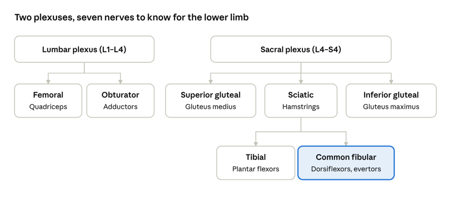
lumbosacral plexus · key nerves and main muscles
The sciatic nerve is really two nerves wrapped together. It splits, usually just above the knee, into the tibial nerve (back of the leg) and the common fibular nerve (front and side of the leg). The common fibular, also called the common peroneal, is highlighted because it's the one most often injured.
The Key Nerves: Where They Get Hurt and What You'll See
Learn each row as a story: this injury damages this nerve, which causes this sign.
|
Nerve (roots) |
Classic injury site |
Main motor loss |
Classic sign |
|
Femoral (L2–L4) |
Pelvic fracture, hip surgery, femoral catheter |
Knee extension (quadriceps) |
Weak knee extension, reduced patellar reflex, numb anterior thigh and medial leg |
|
Obturator (L2–L4) |
Pelvic surgery, difficult childbirth |
Hip adduction |
Weak adduction, numb medial thigh |
|
Superior gluteal (L4–S1) |
Posterior hip dislocation, misplaced gluteal injection |
Hip abduction (gluteus medius and minimus) |
Positive Trendelenburg sign |
|
Inferior gluteal (L5–S2) |
Posterior hip dislocation |
Hip extension (gluteus maximus) |
Trouble climbing stairs or rising from a chair |
|
Sciatic (L4–S3) |
Posterior hip dislocation, misplaced gluteal injection |
Hamstrings and everything below the knee |
Weak knee flexion plus both tibial and fibular deficits |
|
Common fibular (L4–S2) |
Fibular neck fracture, prolonged leg crossing, tight cast |
Dorsiflexion and eversion |
Foot drop, steppage gait, numb dorsum of the foot |
|
Tibial (L4–S3) |
Deep knee trauma; tarsal tunnel at the ankle |
Plantar flexion and inversion |
Can't stand on toes, numb sole of the foot |
Memory hook: PED and TIP. The Peroneal (fibular) nerve Everts and Dorsiflexes, so when it's injured, the foot "dropPED." The Tibial nerve Inverts and Plantarflexes, so when it's injured, you can't stand on your TIPtoes.
L4 vs L5 vs S1: The Root Levels You Must Know
Nerve root questions are where spinal anatomy meets the lower limb. These three levels come up the most, and they're what you'll use in practice every day.
|
Root |
Key motor test |
Reflex |
Sensory area |
|
L4 |
Ankle dorsiflexion (tibialis anterior) |
Patellar |
Medial leg and medial ankle |
|
L5 |
Big toe extension (extensor hallucis longus) |
No reliable reflex |
Top of the foot and first web space |
|
S1 |
Ankle plantar flexion and foot eversion |
Achilles |
Lateral foot and sole |
Connect it to the disc: a posterolateral L4–L5 disc herniation usually compresses the L5 root, and an L5–S1 herniation usually compresses S1. For the reasoning, see our spinal anatomy review.
Spotlight: Foot Drop vs Trendelenburg
These two signs are easy to mix up because both change how someone walks.
- Foot drop (common fibular nerve, or an L5 root problem): the patient can't lift the front of the foot. To keep the toes from catching, they lift the knee high with each step. This is called a steppage gait.
- Trendelenburg sign (superior gluteal nerve): ask the patient to stand on the affected leg. The weak gluteus medius can't hold the pelvis level, so the pelvis drops on the opposite side. The side that drops is the side that's not injured, which is exactly the trap boards like to set.
Test Yourself: 5 Practice Questions
Cover the answers, commit to a choice, then check.
- A patient with a fibular neck fracture can't dorsiflex the ankle and drags the toes when walking. Which nerve is injured?A. Tibial B. Femoral C. Common fibular D. Obturator
- When a patient stands on the right leg, the left side of the pelvis drops. Which nerve is most likely injured? A. Right superior gluteal B. Left superior gluteal C. Right inferior gluteal D. Left femoral
- Which nerve root is tested by the Achilles reflex? A. L3 B. L4 C. L5 D. S1
- A patient has weak big toe extension and numbness on the top of the foot, but normal reflexes. Which root is most likely involved? A. L3 B. L4 C. L5 D. S1
- After hip surgery, a patient has weak knee extension and a reduced patellar reflex. Which nerve is most likely injured? A. Sciatic B. Femoral C. Obturator D. Inferior gluteal
Answers
- The common fibular nerve wraps around the fibular neck. Damage causes foot drop.
- The pelvis drops on the side opposite the weak gluteus medius.
- S1 is the Achilles reflex; L4 is the patellar reflex.
- Big toe extension and the top of the foot point to L5, which has no reliable reflex.
- The femoral nerve supplies the quadriceps and carries the patellar reflex.
Missed one? Add it to your miss log, as we describe in our Part I study plan.
Lower Limb Nerve FAQs
What nerve causes foot drop? Most often the common fibular (common peroneal) nerve, which is easily injured where it wraps around the neck of the fibula. An L5 nerve root problem can also cause foot drop.
What does a positive Trendelenburg sign mean? Weakness of the gluteus medius and minimus, usually from a superior gluteal nerve injury. When the patient stands on the affected leg, the pelvis drops on the opposite side.
What's the difference between the common fibular and common peroneal nerve? Nothing. They're two names for the same nerve. Newer anatomy terms use "fibular," but you'll see both.
What are the L4, L5 and S1 reflexes? L4 is the patellar reflex and S1 is the Achilles reflex. L5 has no reliable reflex, so test it with big toe extension instead.
Where can I review the upper limb? Our brachial plexus review covers the upper limb in the same format.
Master the Anatomy You'll Use Every Day
Lower limb nerves aren't just board material. They're the foundation of every low back and leg exam you'll do as a chiropractor. Learning them well now pays off twice.
The Chiro AlignEd Learning Part I Review Course helps you lock them in:
- Exam-aligned lessons that connect nerves, root levels and clinical signs the way boards test them
- Searchable videos so you can pull up the lumbosacral plexus or any topic in seconds
- Quizzes and full-length practice exams to build fast, confident recall
- Access Until You Pass™ Guarantee: meet our Exam Readiness Standards and if you don't pass, we renew your 90 days of access at no cost
"The review course exceeded my expectations. Everything felt current, clear and thoughtfully organized." — Taylor R., Part I & II Review student
Start the Part I Review Course →
Want more free reviews like this one? Get weekly board exam content in your inbox.
Don't Miss Any Tips!
New tips, tactics and strategies for effective studying and test-taking sent straight to your inbox.
We hate spam. We will never sell your information, for any reason.
