
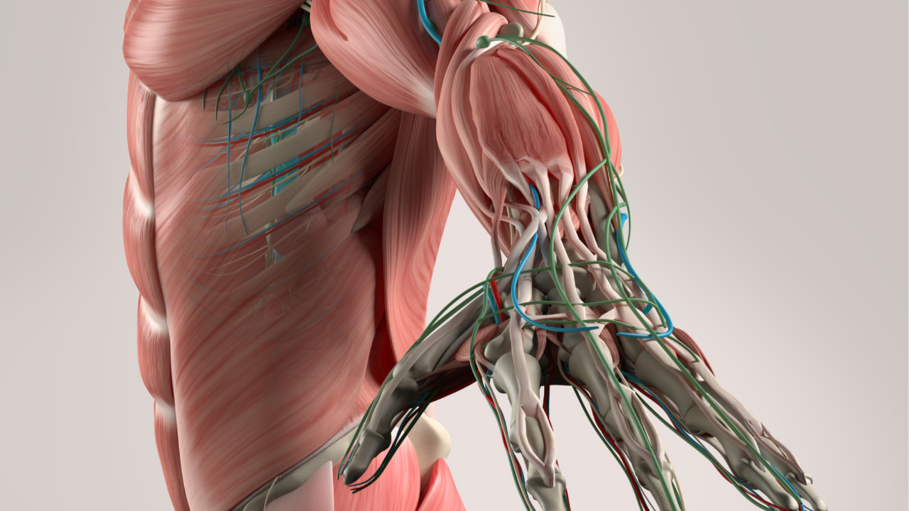
Brachial Plexus Lesions Made Simple: A Board Review for NBCE Part I
Ask any chiropractic student which anatomy topic they dread most, and the brachial plexus is near the top of the list. Five roots, three trunks, six divisions, three cords, a tangle of branches, and a board exam that loves to ask what happens when one of them gets damaged.
Here's the good news: you don't need to memorize the plexus as a picture. You need to know a handful of nerves, where each one gets injured, and the one sign that gives each injury away. That's how the exam asks about it.
This guide breaks it down in that order: the structure, the five terminal nerves, the two classic birth palsies, and practice questions to lock it in.
If you haven't yet, start with our 10-week NBCE Part I study plan. And if you want nerve roots and dermatomes in context, our high-yield spinal anatomy review pairs well with this one.
The Structure in 60 Seconds
The plexus runs in five levels from the spinal cord to the arm. The classic mnemonic gives you the order: Randy Travis Drinks Cold Beer.
brachial plexus · roots to terminal branches
Two shortcuts save you on test day:
- The posterior cord runs the back of the arm. Its two big branches, axillary and radial, supply the deltoid and the extensors.
- The median nerve is the only one built from two cords (lateral and medial). If a question mentions a contribution from both, think median.
The 5 Terminal Nerves: Where They Get Hurt and What You'll See
Most brachial plexus questions give you an injury and ask for the deficit, or give you a deficit and ask for the nerve. Learn each row as a story: this injury damages this nerve, which causes this sign.
|
Nerve (cord) |
Classic injury site |
Main motor loss |
Classic sign |
|
Axillary (posterior) |
Surgical neck of humerus fracture, anterior shoulder dislocation |
Deltoid, teres minor |
Weak abduction past about 15°, numb patch over the lateral shoulder ("regimental badge" area) |
|
Radial (posterior) |
Midshaft humerus fracture (radial groove), crutch pressure in the axilla |
Wrist and finger extensors, triceps (if high) |
Wrist drop |
|
Musculocutaneous (lateral) |
Rarely injured alone; deep upper-arm trauma |
Biceps, brachialis, coracobrachialis |
Weak elbow flexion and supination, numb lateral forearm |
|
Median (lateral + medial) |
Carpal tunnel at the wrist; supracondylar humerus fracture at the elbow |
Thenar muscles; forearm flexors if high |
Thenar wasting ("ape hand"); hand of benedictionwhen a high lesion patient tries to make a fist |
|
Ulnar (medial) |
Medial epicondyle fracture or pressure at the elbow; Guyon's canal at the wrist |
Most intrinsic hand muscles |
Claw hand of the 4th and 5th digits, numb medial hand |
Memory hook: follow the bone. The axillary nerve wraps the surgical neck, the radial nerve spirals around the midshaft, and the ulnar nerve sits behind the medial epicondyle (your "funny bone"). A fracture at each spot points straight to its nerve.
Erb's Palsy vs Klumpke's Palsy
These two trunk injuries are board favorites because the mechanism tells you the answer. Ask yourself: was the neck pulled away from the shoulder, or was the arm pulled up overhead?
|
|
Erb's palsy (upper trunk) |
Klumpke's palsy (lower trunk) |
|
Roots |
C5–C6 |
C8–T1 |
|
Mechanism |
Head and shoulder forced apart (difficult delivery, fall onto the shoulder) |
Arm yanked upward (breech delivery, grabbing a branch while falling) |
|
Muscles affected |
Deltoid, supraspinatus, infraspinatus, biceps |
Intrinsic hand muscles |
|
Classic sign |
"Waiter's tip": arm adducted, medially rotated, elbow extended, forearm pronated |
Total claw hand |
|
Extra clue |
— |
Possible Horner syndrome if the T1 sympathetic fibers are involved |
A cervical rib or tight scalene muscles can compress the lower trunk too, which is why thoracic outlet syndrome often looks like a mild Klumpke picture.
Branches That Come Off Early
A few nerves leave the plexus before the cords, so injuries to them spare the rest of the arm.
- Long thoracic nerve (C5–C7 roots) → serratus anterior. Damage causes a winged scapula, classically after mastectomy or a stab wound to the lateral chest.
- Dorsal scapular nerve (C5 root) → rhomboids and levator scapulae.
- Suprascapular nerve (upper trunk) → supraspinatus and infraspinatus. Damage weakens the start of abduction and external rotation.
Test Yourself: 4 Practice Questions
Cover the answers, commit to a choice, then check.
- A patient with a midshaft humerus fracture can't extend the wrist. Which nerve is most likely injured? A. Median B. Ulnar C. Radial D. Axillary
- A newborn after a difficult delivery holds the arm adducted and medially rotated, with the forearm pronated. Which roots are involved? A. C5–C6 B. C7 C. C8–T1 D. C5–T1
- After a mastectomy, a patient's scapula protrudes when she pushes against a wall. Which nerve was injured? A. Dorsal scapular B. Long thoracic C. Suprascapular D. Thoracodorsal
- A fall onto the elbow fractures the medial epicondyle. Which finding is most likely? A. Wrist drop B. Clawing of the 4th and 5th digits C. Loss of shoulder abduction D. Weak elbow flexion
Answers
- The radial nerve runs in the radial groove at the midshaft. Wrist drop is its signature.
- This is the "waiter's tip" of Erb's palsy, an upper trunk (C5–C6) injury.
- The long thoracic nerve supplies serratus anterior, which holds the scapula against the chest wall.
- The ulnar nerve passes behind the medial epicondyle. Losing its intrinsic hand muscles causes the claw.
Missed one? Add it to your miss log, as we describe in our Part I study plan.
Brachial Plexus FAQs
What's the easiest way to memorize the brachial plexus? Learn the order with "Randy Travis Drinks Cold Beer," then focus on the five terminal nerves. For each one, know where it's injured and its classic sign.
What causes wrist drop? Radial nerve injury, most often from a midshaft humerus fracture or prolonged pressure in the axilla, like improper crutch use.
What's the difference between Erb's palsy and Klumpke's palsy? Erb's palsy affects the upper trunk (C5–C6) and causes the "waiter's tip" posture. Klumpke's palsy affects the lower trunk (C8–T1) and causes a claw hand.
Which nerve causes a winged scapula? The long thoracic nerve, which supplies serratus anterior.
Is the brachial plexus tested on NBCE Part I? Upper extremity nerves and their injuries are core anatomy content, so expect to see them. Check the current NBCE Part I test plan for the official domain breakdown.
Turn Your Toughest Topics Into Easy Points
If the brachial plexus just went from dreaded to doable in one read, imagine that across all six Part I domains.
That's what the Chiro AlignEd Learning Part I Review Course is built to do:
- Exam-aligned lessons that teach high-yield topics the way the NBCE asks about them
- Searchable videos so you can pull up the brachial plexus, or any topic, in seconds
- Quizzes and full-length practice exams to train timing, stamina and decision-making
- Access Until You Pass™ Guarantee: if you don't pass, we renew your 90 days of access at no cost when you meet our Exam Readiness Standards
"What stood out most was how familiar the exam felt." — Hillary W., Part I Review student
Start the Part I Review Course →
Want more free reviews like this one? Get weekly board exam content in your inbox.
Don't Miss Any Tips!
New tips, tactics and strategies for effective studying and test-taking sent straight to your inbox.
We hate spam. We will never sell your information, for any reason.
